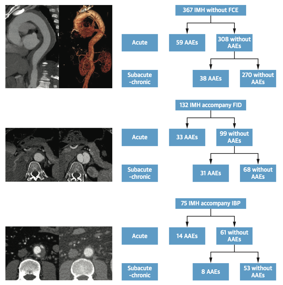
Type B intermural hematoma of the aorta is a common acute aortic syndrome and is often considered a specific type of aortic coarctation, accounting for about 20% of acute aortic syndromes. The hematoma usually forms as a result of rupture of a trophoblastic vessel in the middle layer of the aortic wall and bleeding. Patients experience severe tearing pain in the chest and back at the onset. Conventional wisdom is that this disease is less fatal and conservative treatment with medications is usually recommended. However, an increasing number of case reports have shown that intermural hematomas of the aorta have a 47% probability of progressing to aortic coarctation and a 45% risk of causing aortic rupture, with only about 10% of patients improving with conservative treatment. This makes the previous strategy of conservative treatment with drugs unreasonable. How to determine the prognosis of type B aortic intermural hematoma early enough to avoid unnecessary surgery without missing potentially life-threatening high-risk patients has become an important clinical question that needs to be answered urgently, and there is a lack of relevant clinical studies. On January 23, 2024, Prof. Weiguo Fu and Wisdom Dong of Zhongshan Hospital/Fudan University Institute of Vascular Surgery, Fudan University, led by Dr. Xiaolang Jiang and Dr. Tianyue Pan, published their research results entitled “Prognostic Implications of Initial Focal Contrast Enhancement in Acute Type B Intramural Hematoma” online in the Journal of the American College of Cardiology, the leading journal in the field of cardiovascular diseases (IF 24), which is a publication of the American Heart Association. The study entitled “Prognostic Implications of Initial Focal Contrast Enhancement in Acute Type B Intramural Hematoma” was published online in the Journal of the American College of Cardiology (IF 24), a prestigious cardiovascular journal of the American College of Cardiology and a journal of the American Heart Association. Through in-depth analysis of more than 500 cases of type B intramural hematoma in the past 10 years, the study identified the important indicator of initial Focal Contrast Enhancement (FCE) in the aortic wall as a predictor of the development of type B intramural hematoma. For the first time, the evolution of FCE in the natural course of aortic intermural hematoma has been systematically described, and the corresponding correlation between different types and locations of FCE and the prognosis of aortic intermural hematoma has been subdivided, which provides a key indicator for predicting the progression of aortic intermural hematoma, and accordingly, a new strategy of aortic intermural hematoma treatment has been put forward, which provides an important guideline for the differentiation of the aortic intermural hematoma in the clinical setting, and the rational management of the aortic intermural hematoma. In this study, approved by the Ethics Committee of Zhongshan Hospital of Fudan University and registered in the China Clinical Trial Center, 574 type B aortic intermural hematomas were included from 3259 acute aortic syndromes from 2009 to 2019, which were classified into the group with FCE (n=207) and without FCE (n=367) according to the immediate thoracic and abdominal aortic CTA results. Among them, the FCE group was typed into subgroups of focal intimal dissection (FID, n=132), which were subintimal contrast-enhancing foci communicating with the aorta with a dissection >3 mm, and intramural blood pools (IBP, n=75), which were subintimal contrast-enhancing foci with an intact intima or a dissection ≤3 mm. These patients were treated with standard conservative therapy and close follow-up with CTA. The primary study endpoints were all-cause death, aortic-related death, aortic complications requiring surgical intervention, and aortic-related adverse events. The median follow-up for all patients was 42 months, 48 months in the FCE group and 36 months in the non-FCE group. The findings suggest that type B aortic intermural hematomas with combined FCE were more likely to develop adverse aortic events, with a 5-year freedom-from-intervention rate of 59.7%, compared with a 5-year freedom-from-intervention rate of 73.3% in the group without combined FCE. Multifactorial Cox regression analysis also confirmed FCE as an independent risk factor for aortic adverse events in patients with aortic intermural hematoma, with pleural effusion being the other factor. This finding alerts clinicians to the necessity of observing FCE in the first CTA at the acute onset of aortic intermural hematoma! In subgroup analyses, 25% of patients in the FID group and 18.7% of patients in the IBP group progressed in the acute phase, and 34.3% of patients in the FID group and 13.3% of patients in the IBP group developed adverse aortic events in the subacute-on-chronic phase.The 5-year freedom-from-intervention rate was 50.8% in the FID group, which was significantly lower than that in the IBP group (75.3%).Adverse aortic events occurred in the FID group in population, approximately 65.6% of the ruptures were located in the proximal aorta; whereas in the population with combined IBP, the occurrence of adverse aortic events was not related to location. Further multifactorial Cox regression analysis also confirmed that FID in the proximal aorta was an independent risk factor for aortic adverse events, whereas IBP was not. Summarizing the above studies, it can be found that patients with FID on initial CTA are prone to aortic adverse events in both acute and subacute-chronic phases, especially those with rupture located in the proximal aorta are more prone to progression, therefore, long-term close follow-up is needed if conservative treatment is chosen, and surgical interventions are preferred if there is progression on imaging or symptomatic recurrence. For patients with IBP, close follow-up is also required during the acute and subacute phases, with less frequent follow-up when the patient enters the chronic phase. In conclusion, this study is the first to systematically elucidate the evolution of FCE as an imaging feature in the natural course of type B aortic intermural hematoma, confirms through a large sample size that the observation of FCE typing and location in the acute phase of aortic intermural hematoma directly affects the prognosis of the patients, reveals that FID located in the proximal part of the aorta is a key factor influencing the aortic adverse events, and offers new perspectives on prevention and treatment and follow-up strategies of type B aortic intermural hematomas. strategy provides a new perspective.


