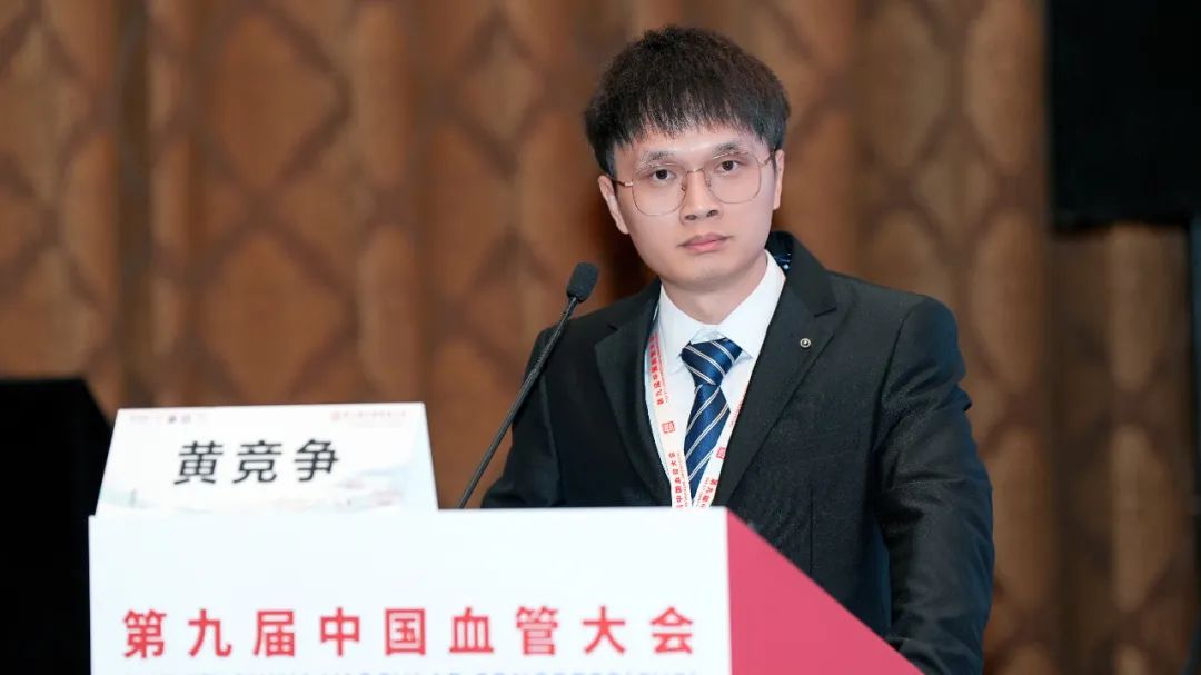
During the 9th China Vascular Conference (CVC 2024), held on September 12, 2024, at the Chengdu Century City International Convention Center, the annual Top 10 Case Showcase of the Vascular Surgery Group of the Chinese Clinical Case Database was successfully held. Numerous surgical experts provided in-depth analysis and experience sharing on the selected top 10 cases. Professor Huang Jingzheng from the Department of Vascular Surgery, Second Affiliated Hospital of Nanchang University, shared a case of endovascular treatment for a giant aortic arch aneurysm with severe tortuosity of the thoracic aorta.

Case Sharing
Patient Information (Female, 61 years old)
Chief Complaint: Aortic arch aneurysm detected one day ago during an examination.
Current Medical History: One day ago, the patient underwent a CT examination at an outside hospital and was diagnosed with an aortic arch aneurysm. The patient reported no particular discomfort and denied any chest tightness, shortness of breath, chest pain, dizziness, headache, nausea, or vomiting.
Past Medical History: Denied history of hypertension, diabetes, or coronary heart disease.
Physical Examination: Conscious, clear lung sounds, regular heart rhythm, no murmurs in any valve areas, and normal limb movements.
Preoperative CTA: Giant aortic arch aneurysm with a diameter of 11 cm, severe tortuosity of the thoracic aorta, compression of the left common carotid artery (LCCA) and left subclavian artery (LSA).

Case Characteristics and Challenges
• Lesion Characteristics: Giant aortic arch aneurysm with a maximum transverse diameter of 110 mm, severe thoracic aorta tortuosity. The aneurysm compressed the arch branches, causing a narrowed LCCA.
• Surgical Challenges: Open thoracotomy would be highly invasive with a long surgery duration; exposure would be challenging due to the large aneurysm, and there is a high risk of rupture during surgery.
• Endovascular Challenges:
• Path Establishment: How to deliver the stent to the proximal anchoring zone through the severely tortuous thoracic aorta?
• Proximal Anchoring Zone Selection: Requires extending the anchoring zone to the posterior margin of the innominate artery (IA).
• Distal Anchoring Zone Selection: Should it extend beyond the curve?
• Stent Selection: There is significant diameter variation (27.6 mm–15.5 mm) between the IA and the anchoring zone within the aneurysm. How should the oversize be chosen? What about stent compliance and wall apposition?
• Arch Branch Reconstruction: Should surgical intervention or endovascular techniques be used?
Surgical Plan and Device Selection
Considering all factors, endovascular intervention was chosen. Using a traction pathway and the double-sheath kissing technique, the stent was delivered to the posterior margin of the IA. The staged release technique was used for precise positioning, and proximal angle control ensured complete apposition of the stent on the lesser curvature of the aorta.
Management of Arch Branches:
• LCCA Reconstruction: Decided between in situ fenestration or chimney technique.
• LSA Occlusion: Considered for closure.
Device Selection:
• Aortic Stent: A controllable covered thoracic aortic stent.
• Hydrophilic-Coated Guide Sheath.
Surgical Procedure
1. Right Femoral and Right Axillary Artery Approaches: Vascular sheaths were inserted, and routine pigtail catheter angiography was performed.

2.Right Axillary Artery Approach: The guidewire was captured in the aortic arch, establishing a traction pathway.

3.Right Femoral Artery Approach: The hydrophilic-coated sheath was advanced to the proximal thoracic aorta using traction techniques.

4.Right Axillary Artery Approach: Another hydrophilic-coated sheath was advanced, aligning the two sheaths for pathway establishment.

5.Right Femoral Artery Approach: The controllable covered thoracic aortic stent was introduced. As the stent was advanced, the axillary sheath was withdrawn to position the stent at the proximal anchoring zone, just beyond the IA.

6.Angiography: Confirmed that the stent was successfully positioned at the IA’s posterior margin, and a pathway was established for the LCCA.

7.Stent Release: The stent was opened from the proximal end to the middle diameter. Angiography showed ideal positioning.

8.Full Stent Deployment: The stent was released to its full diameter. Angiography showed a minor endoleak, which was reduced by adjusting the proximal angle for better wall apposition.

9.LCCA Fenestration Attempts: Multiple attempts were made for in situ fenestration, but due to the sharp angle of the LCCA, fenestration was challenging.

10.LCCA Reconstruction via Chimney Technique: The chimney technique was used for LCCA reconstruction. Angiography confirmed good results with unobstructed blood flow.

11.LSA Occlusion: LSA was occluded via the left brachial artery approach.

Final Angiography: The aneurysm was no longer visualized, LCCA flow was unobstructed, and LSA occlusion was successful. Cerebral angiography confirmed unobstructed flow in the arch branches and intracranial circulation.

Postoperative Follow-Up
Thoracic aortic imaging showed good stent visibility, complete thrombosis of the aneurysmal sac, and patent grafts without endoleaks, dislocation, thrombosis, or stenosis. The proximal stent showed no “bird beak” sign.

Conclusion
Endovascular treatment using a compliant, controllable thoracic aortic covered stent system for severely tortuous giant aortic arch aneurysms is safe and effective. Establishing a pathway using traction guidewires and the double-sheath kissing technique allows for smooth stent delivery through a severely tortuous thoracic aorta. The management of arch branches should be tailored to the patient’s anatomical characteristics and tolerability, developing different strategies accordingly.


