The 2nd Tianfu Vascular Conference (TFS 2024) was successfully held on May 17-18, 2024, in Chengdu. From the pinnacle of medical science, we foresee the future of the vascular discipline, recognizing the immense responsibility and honorable mission ahead. As members of the vascular field, we are tasked with advancing the discipline and improving diagnostic and treatment standards. In the future, we will analyze the content of TFS lectures to help you understand the most cutting-edge vascular treatment experiences.
In this series of articles, we will thoroughly analyze the presentation by Professors Yang Baihui and Yin Chengyong from the Second Affiliated Hospital of Kunming Medical University at the Tianfu Vascular Conference. They discussed their experiences and cases in using multiple techniques to treat complex diseases involving major abdominal vessels.

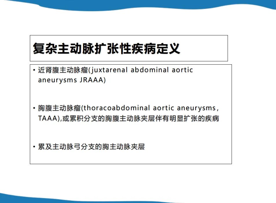
Definition of Complex Aortic Aneurysms
• Juxtarenal Abdominal Aortic Aneurysms (JRAAA)
• Thoracoabdominal Aortic Aneurysms (TAAA): Aneurysms involving the thoracoabdominal aorta with significant dilation and dissection extending to branches.
• Aortic Dissections Involving the Aortic Arch Branches
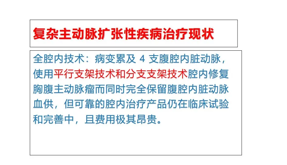
Current Treatment Approaches
1. Total Endovascular Techniques: Using parallel stent technology and branched stent technology for endovascular repair of thoracoabdominal aortic aneurysms, preserving visceral arterial supply. However, these techniques are costly and still in clinical trial stages.
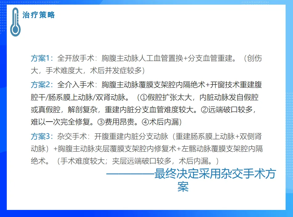
2. Hybrid Surgery: Combining visceral arterial debranching surgery with thoracoabdominal aortic covered stent endovascular repair reduces the significant trauma and high complication rates of traditional surgery, avoiding the high endoleak rates and costs of total endovascular treatment.

Case Studies
Case 1:
• Patient: Male, 57 years old, admitted for back pain lasting one week. He had undergone Bentall surgery for ascending aortic dissection aneurysm 16 years ago. Recent severe back and abdominal pain prompted a CT scan, which revealed a descending aortic dissection with a large abdominal aortic aneurysm.
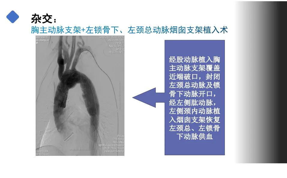
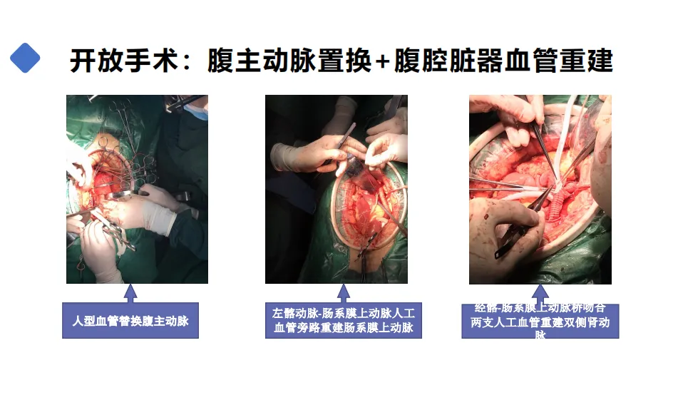
• Surgical Method: Hybrid technique, abdominal aortic aneurysm graft replacement, visceral artery reconstruction, thoracic aortic stent, and left subclavian artery-left common carotid artery chimney stent implantation.

• Surgical Process:
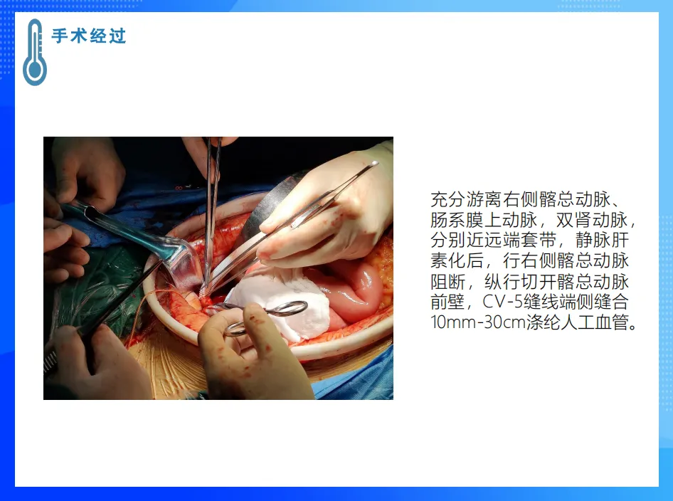
• Entering the retroperitoneal space via a midline abdominal incision around the navel, an aneurysmal expansion of the abdominal aorta was found. The right renal artery received blood from the true lumen, while the left renal artery was supplied by the false lumen.
• The right common iliac artery was clamped, and the anterior wall of the common iliac artery was incised for end-to-side anastomosis with the graft.
• The graft was reconstructed with the superior mesenteric artery and bilateral renal arteries, ensuring good postoperative blood flow.
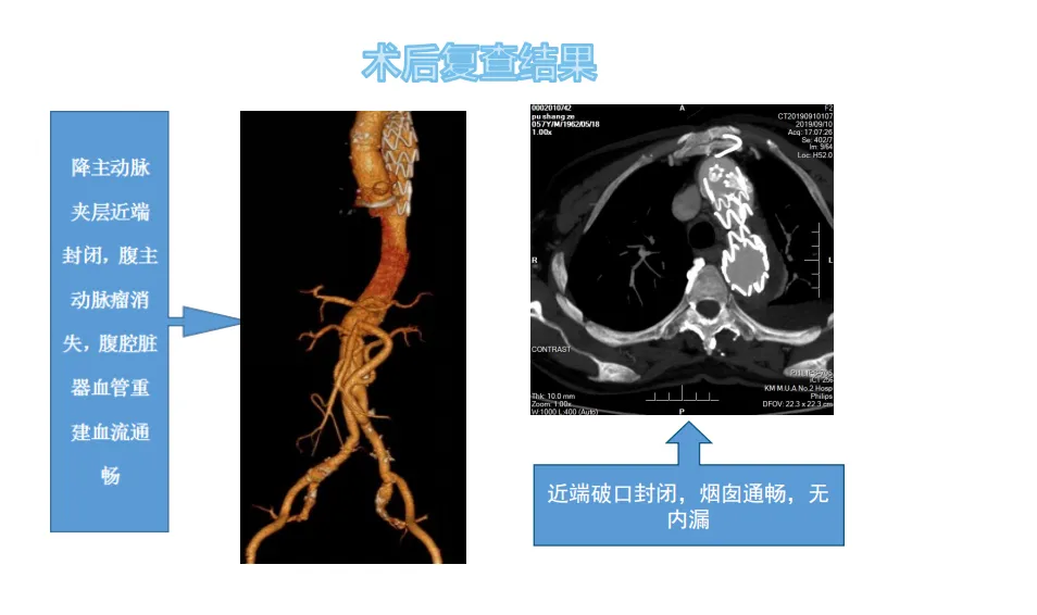
• Postoperative Results: CT showed good recovery of the true lumen and thrombosis of the false lumen.
Case 2:
• Patient: Male, 44 years old, admitted for left-sided chest and abdominal pain lasting 20 days. He had previously undergone endovascular exclusion for thoracic aortic dissection nine years ago. CT showed thoracoabdominal aortic dissection with aneurysmal expansion and thrombus formation around the false lumen.
• Surgical Method: Hybrid surgery – open reconstruction of visceral branch arteries (reconstructing the superior mesenteric artery and bilateral renal arteries) and thoracoabdominal aortic dissection covered stent endovascular repair, with left iliac artery covered stent exclusion.
• Surgical Process:
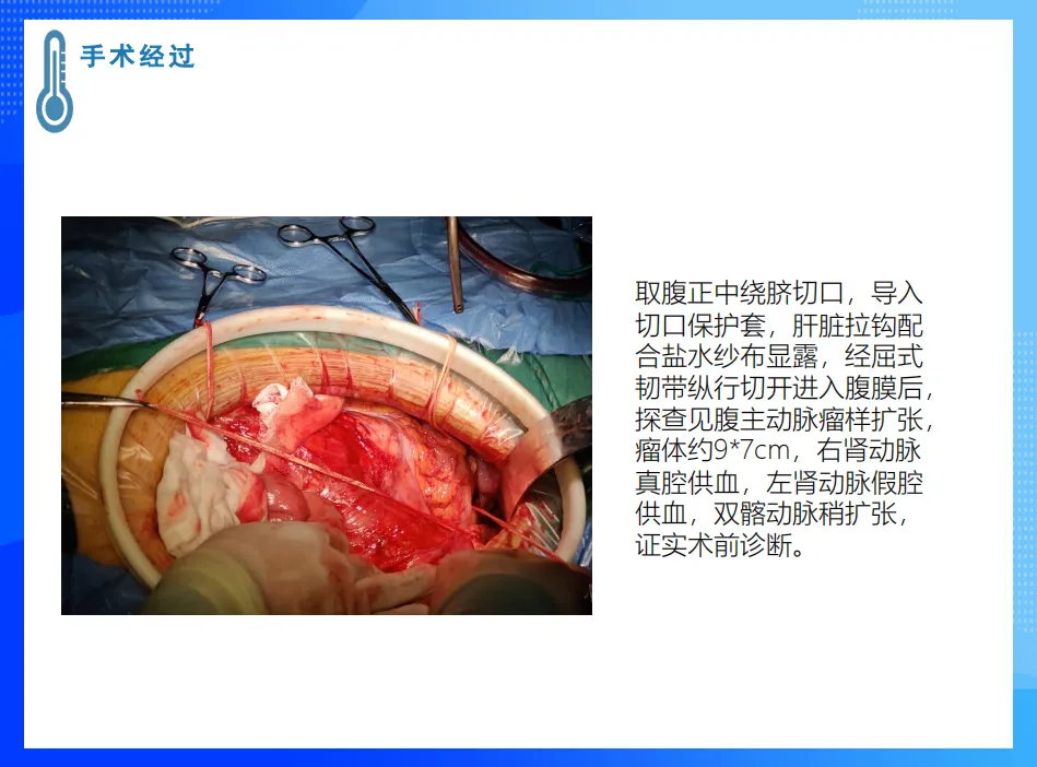
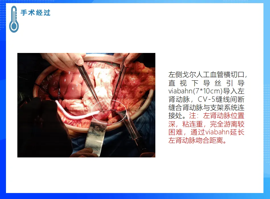
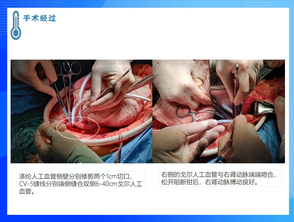
• Opening the abdomen revealed aneurysmal expansion of the abdominal aorta. The right renal artery was supplied by the true lumen, while the left renal artery was supplied by the false lumen.
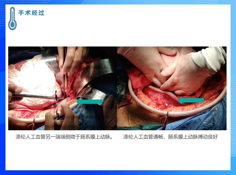
• Reconstruction of the superior mesenteric artery and bilateral renal arteries ensured restored blood flow.
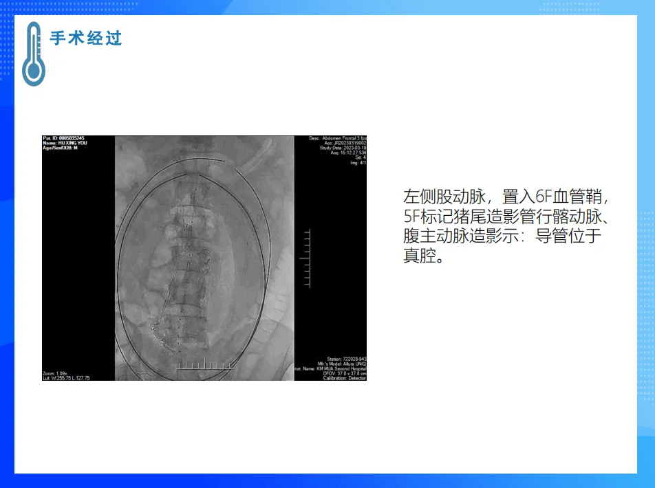
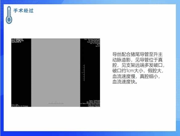
• A thoracic aortic stent was implanted via the femoral artery to cover the proximal tear and seal the left common carotid and subclavian artery openings.
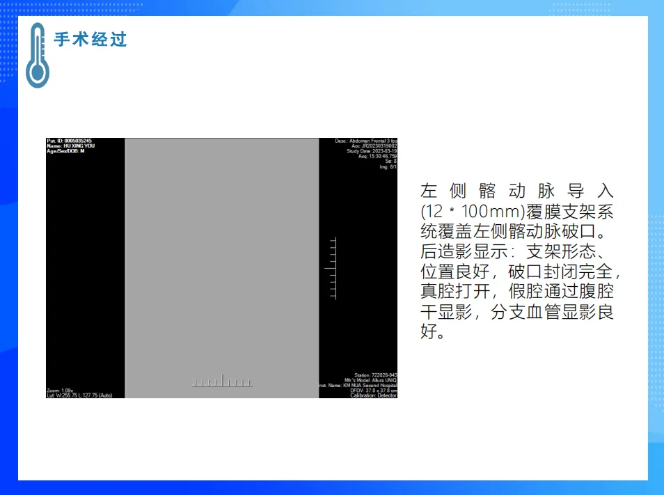
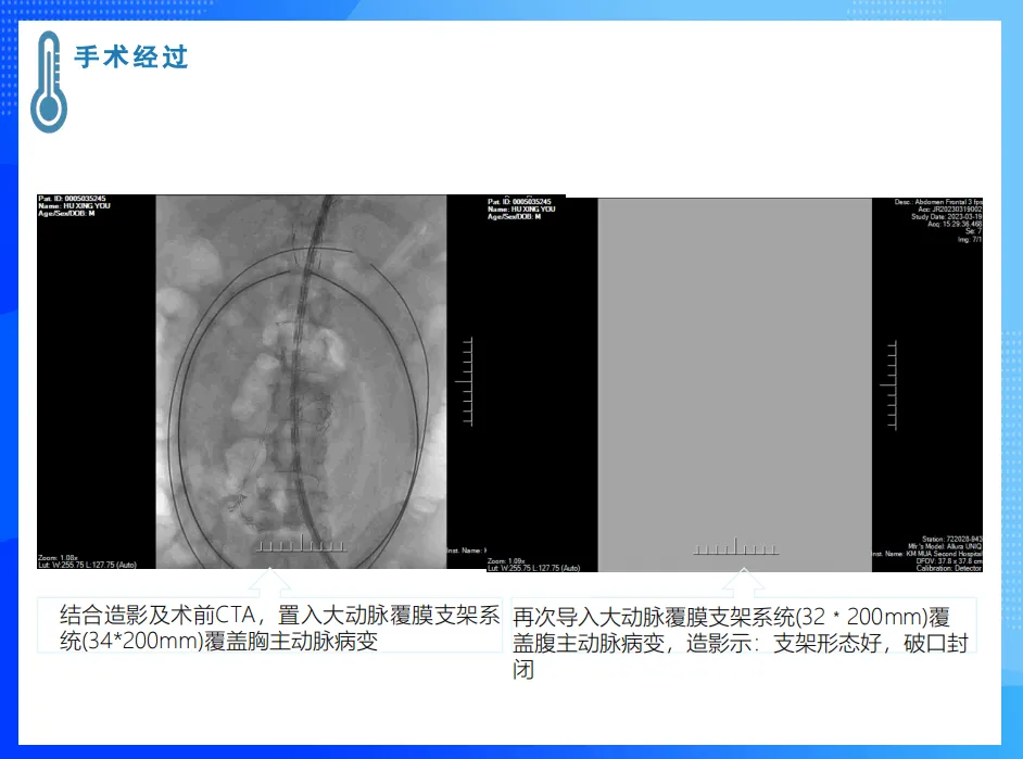
• Postoperative Results: Follow-up showed good stent morphology, sealed tears, and normal recovery of the true lumen.
Case 3:
• Patient: Female, 49 years old, admitted for a retroperitoneal mass with fatigue lasting one year. CT indicated multiple hypervascular masses in the retroperitoneal and pelvic areas, involving the uterus, left adnexa, and part of the left colon.
• Surgical Method: Hysterectomy with bilateral adnexectomy, removal of the inferior vena cava tumor, reconstruction of the inferior vena cava, and exploration of the intestines and bilateral renal veins.
• Surgical Process:
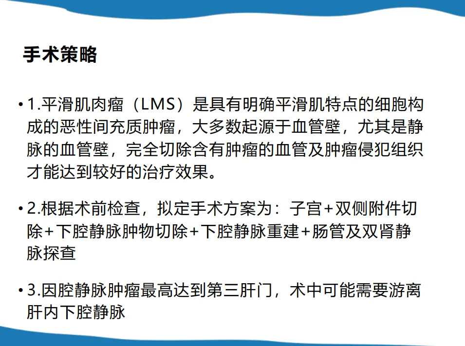
• Exploratory laparotomy revealed the tumor invading part of the sigmoid colon, which was resected along with the involved colon segment and anastomosed.
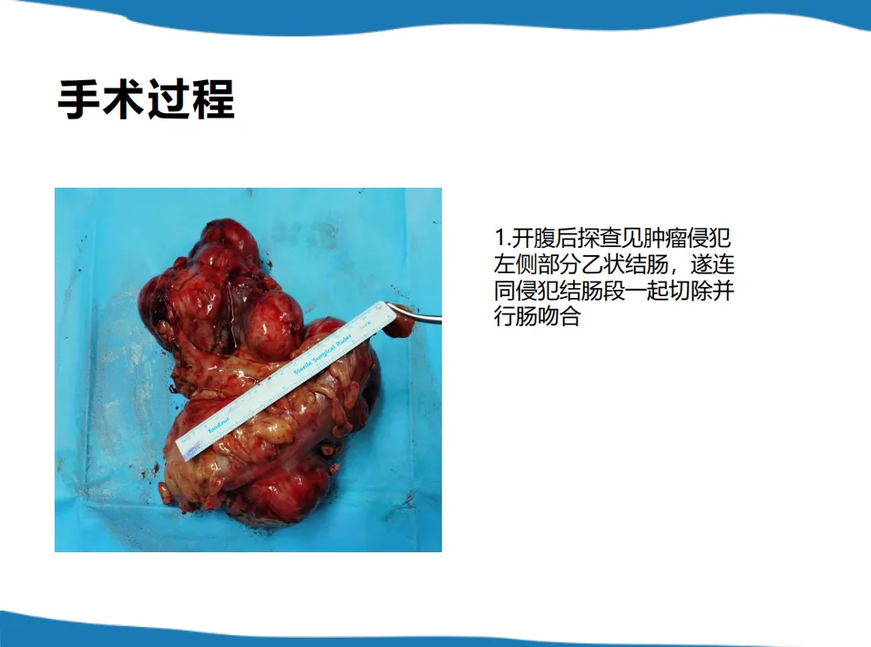
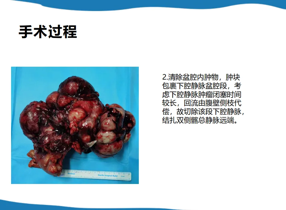
• Pelvic mass removal involved excision of the inferior vena cava pelvic segment.
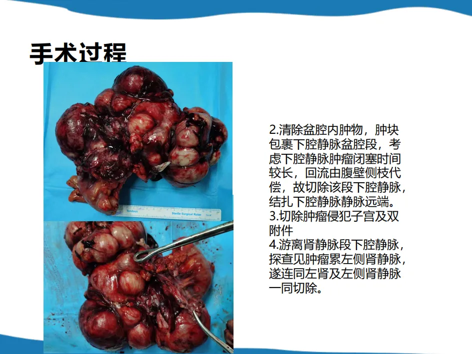
• The uterus and bilateral adnexa were resected, and the inferior vena cava segment involving the renal veins was freed, revealing tumor involvement of the left renal vein, which was resected along with the left kidney and left renal vein.
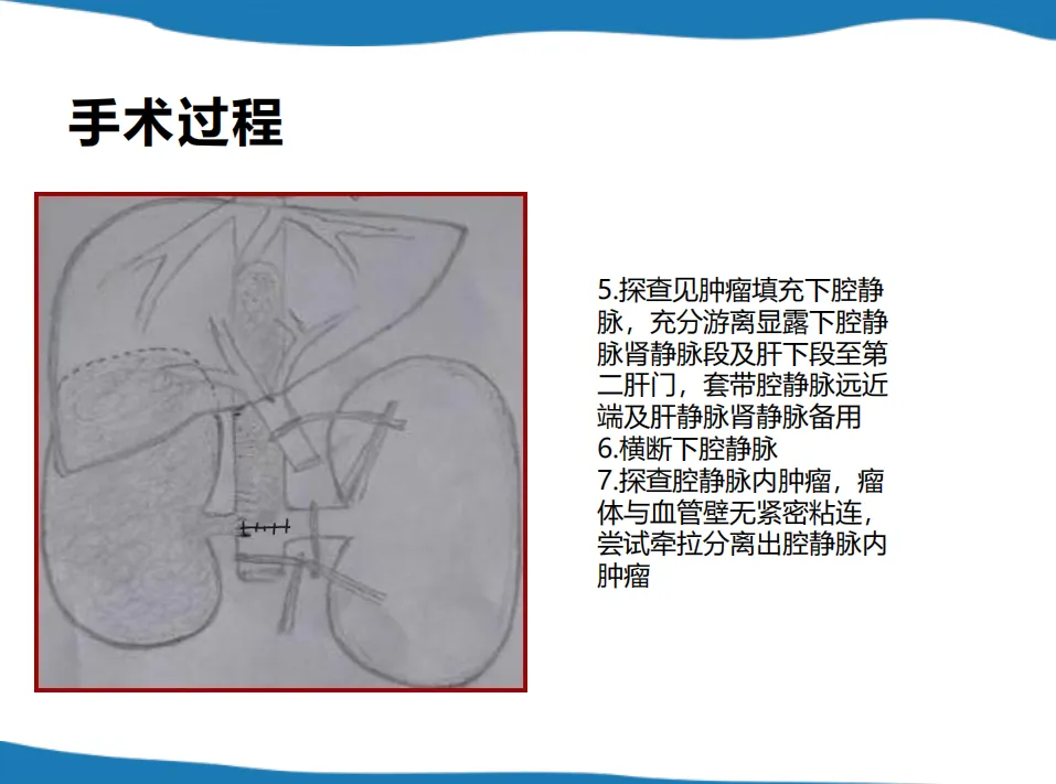
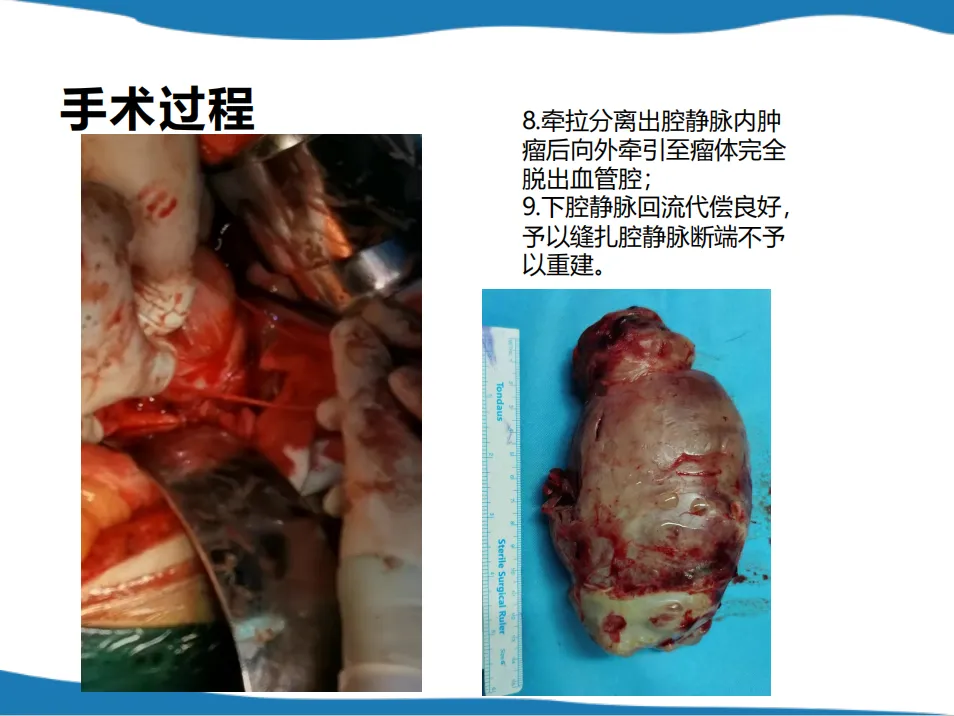
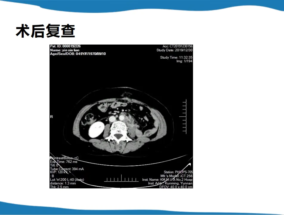
• Postoperative Results: CT showed complete tumor resection with good patient recovery.
Summary and Outlook
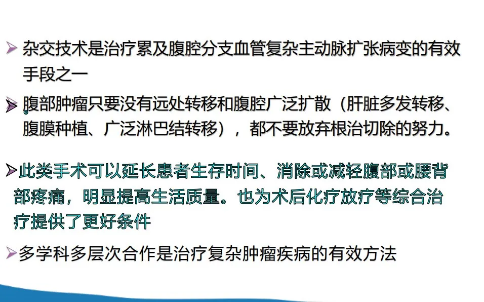
Hybrid techniques are effective for treating complex aortic expansions involving abdominal branches. For abdominal tumors without distant metastasis or extensive abdominal spread, efforts should be made for radical resection. Multidisciplinary and multi-tiered collaboration is an effective approach to treating complex tumor diseases.
Contact Us
In upcoming articles, we will continue to provide more in-depth analysis on treating complex abdominal vascular diseases. Stay tuned! If you have any questions or interests regarding the treatment of complex abdominal vascular diseases or the Tianfu Vascular Conference, please leave a comment or contact us via email at endovascluar@simtomax.cn. Thank you for your attention. Let’s work together for health!


