
Author: Dr. R. de Graaf
Institution: Department of Interventional Radiology, Clinic of Friedrichshafen, Germany
Summary
This presentation explores the diagnostic value of imaging in pelvic congestion syndrome (PCS), highlighting the application of MR venography (MRV), angiography, and intravascular ultrasound (IVUS) in diagnosing PCS. The study shows that non-invasive imaging can effectively identify venous-origin lesions, while angiography and IVUS offer detailed insights into blood flow patterns and lesions, particularly when simultaneous treatment is needed.
Key Aspects of Imaging Diagnosis for PCS
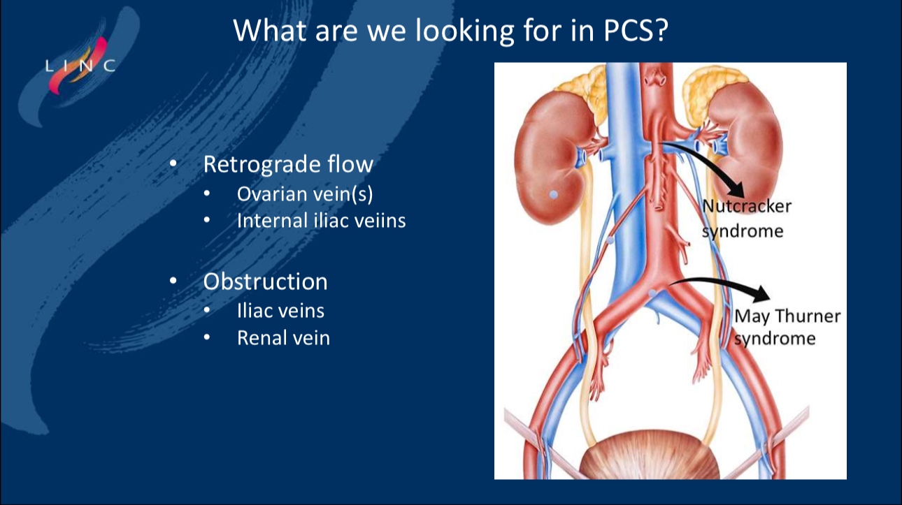
• Key Diagnostic Points: Imaging for PCS focuses on detecting the following:
• Retrograde Blood Flow: Especially in the ovarian veins and internal iliac veins.
• Venous Obstruction: Primarily focusing on narrowing of the iliac and renal veins.
Imaging Diagnostic Methods
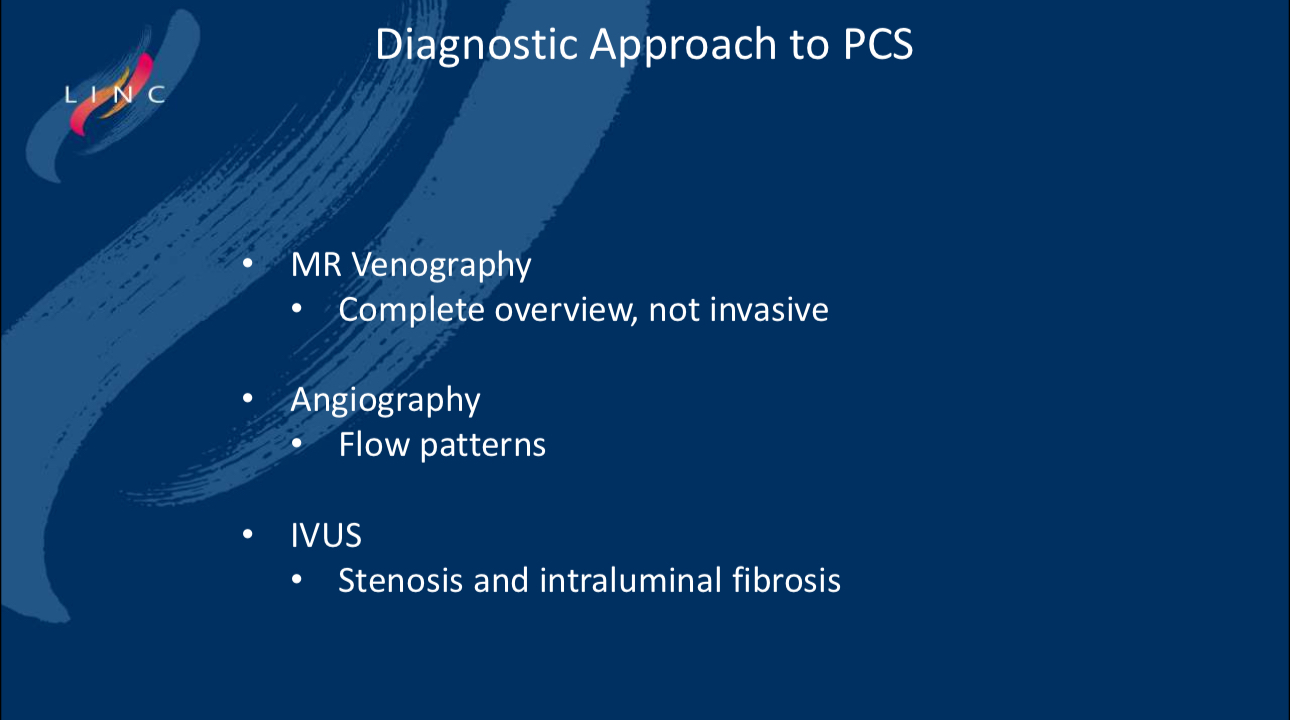
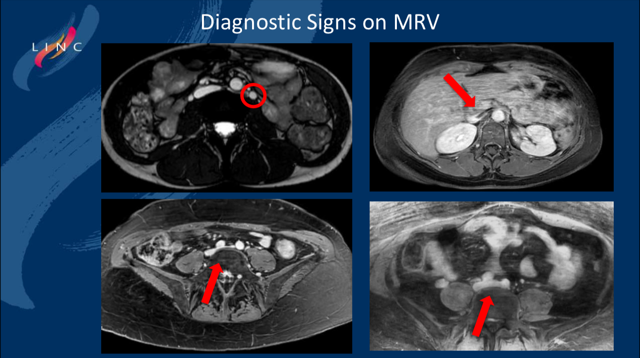
• MR Venography (MRV): MRV, as a non-invasive imaging technique, provides a comprehensive view of the pelvic veins and is crucial for excluding other causes, especially when results are negative. MRV offers a complete anatomical view without invasive harm to the patient.
• Angiography and IVUS: Angiography helps identify venous lesions by observing blood flow patterns. IVUS provides a more detailed assessment of the degree of narrowing and fibrosis within the lumen, making it valuable in diagnosing stenotic or obstructive lesions and can be performed concurrently with treatment.
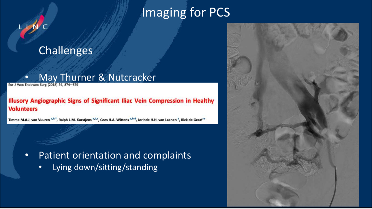
Clinical Challenges
• Impact of Body Position: There are diagnostic challenges in PCS, such as the patient’s body position affecting diagnostic results. Symptoms and venous blood flow patterns differ when patients are lying down, sitting, or standing, so these factors must be fully considered during diagnosis.
Conclusion
1. Imaging plays a crucial role in identifying venous lesions in PCS, particularly providing key information before diagnostic and therapeutic interventions.
2. MRV, due to its non-invasive nature, is highly effective in initial screenings, while angiography and IVUS offer more in-depth diagnostic information, especially when combined with treatment.
3. Future diagnostic protocols should incorporate multiple imaging techniques to ensure optimal diagnostic accuracy and patient outcomes.
Contact Us
• Email: endovascluar@simtomax.cn
More international information available at:
• Facebook: Vasco Knight
• Instagram: knight_vasco
Let’s safeguard health together and showcase your brilliance to the world!